Characterising the gut nervous system in a disease mouse model
Cellular and molecular profiling in preparation for stem cell transplant therapies
Highlights
- Microscopy confocal microscopy
- Dissection preparation of histological sections
- Immunohistochemistry labelling samples with antibodies to detect the presence and abundance of certain proteins
- qPCR quantitative technique for measuring gene expression levels in tissues
Overview
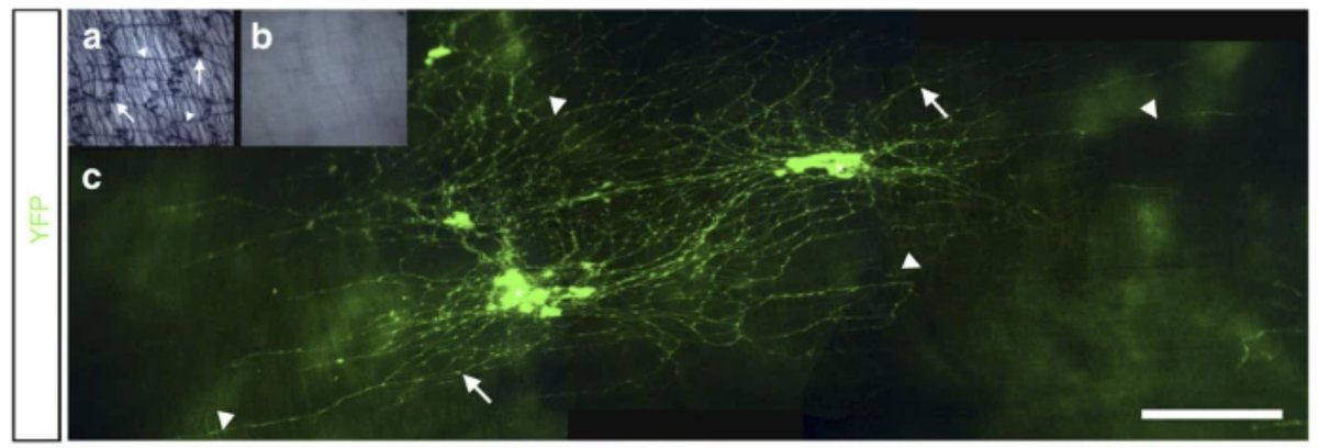
During my master’s degree, I worked in the group of Dr Alan Burns under the supervision of Dr Conor McCann. Dr McCann’s research centred on determining the feasibility of transplanting enteric neural stem cells (ENSCs) into the gastrointestinal system of a mouse model of human enteric neuropathy (nNOS-/-) to rescue the impaired gut motility observed in this mouse model.
I was specifically involved in characterising the populations of cells within the enteric nervous system of these nNOS-/- mice at baseline to have a reference against which to compare the populations found after the ENSC transplants. Among other things, I was introduced to the technique of quantitative polymerase chain reaction (qPCR) to measure gene expression levels in tissues and performed dissections of enteric tissues and immunohistological stainings to uncover the levels of certain proteins present in this tissue.
Links
Acknowledgements
This project was carried out under the supervision of Dr Conor McCann, then a postdoctoral researcher in Dr Alan Burns’ group at the UCL and Great Ormond Street Institute of Child Health.