Responses to visual threats in mouse models of autism
How genes linked to autism affect a circuit that drives innate responses to visual threats
Highlights
- Built an end-to-end experimental pipeline in MATLAB, from visual stimulus presentation through to analysis of behavioural and electrophysiological recordings.
- Designed and ran an independent, multi-year research project, published as first author.
- Performed stereotaxic surgery in mice to implant optic fibres and silicon probes and to deliver viral constructs.
- Initiated and led cross-group collaborations beyond my immediate team.
- Mentored students at graduate, undergraduate and high-school level alongside the research.
My PhD project investigated whether altered functioning in a subcortical visual pathway could be responsible for some of the attentional differences observed in mouse models of autism.
Background
Autism has a complex and unknown aetiology. What is known however, is that it is not caused by a single mutation in a single gene, instead it is polygenic, with differences in hundreds, or thousands, of genes likely interacting to results in the physiological and behvaioural differences observed in autistic individuals. Disruptive variants of certain genes, like SETD5, however have been found with significantly increased frequencies among autistic individuals and therefore have been classified as autism “risk genes”. These risk genes serve as valuable experimental starting points to probe the potential biological consequences of these genetic differences.
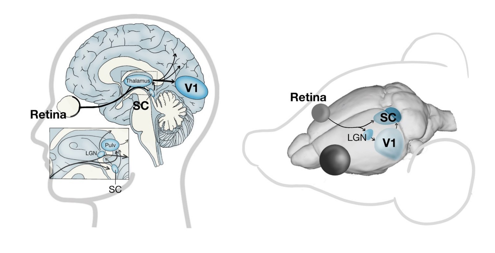
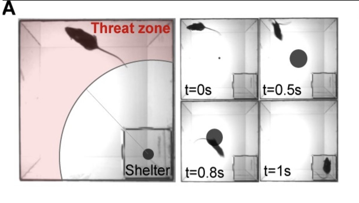
While researching the differences in visual attention and overall sensory differences in autism, I was intrigued to see that little autism research investigated the circuits driving subconscious visual processing through subcortical circuits, especially through a brain region called the superior colliculus (SC), and instead focussed on the predominant visual processing pathway through the lateral geniculate nucleus and onto the visual cortex. The SC is known to be critical in mediating rapid, visually-evoked responses such as escape responses to looming visual stimuli (dark, expanding dots that mimic the approach of an aerial predator).
Hypothesis I
I speculated whether visual processing through this subcortical visual pathway might be affected in the Setd5 mouse model of autism.
NOTE:
Mice lacking one, functional copy of the gene will be referred to as Setd5+/-, whereas their wild-type littermates will be referred to as Setd5+/+. Given a single litter would normally have a mixture of +/- and +/+ pups, we always tested sex-matched, sibling pairs together to ensure that the mice are as comparable as possible.
Methods & Results
To test my hypothesis I investigated both the behavioural and physiological differences of these mice.
- I rigorously analysed the behavioural responses to the looming stimulus.
- I recorded the electrophysiological responses of neurons within the SC, that receive direct visual information from the retinal ganglion cells within the eye, to visual stimuli in order to ascertain if mutations in Setd5 result in fundamental differences in SC visual processing.
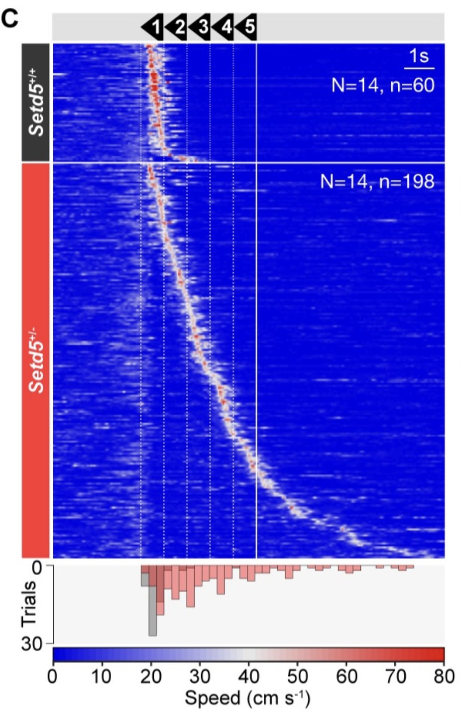
From these experiments, I found that although the Setd5+/- mice were slower at initiating an escape response, I couldn’t identify any major differences in their visual responses within the SC.
With this is mind, I went back to what was known about this circuit. As Evans et al. 2018 described, this neural circuit functioned like a drift diffusion model. Cells within the underlying brain region to the SC, called the periaqueductal grey (PAG), must integrate the input they receive from SC cells, as well as others, and only when their membrane has become sufficiently depolarised will they start firing themselves and signal to downstream motor regions to trigger an escape response.
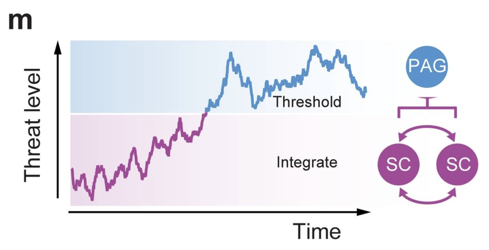
This led us to reconsider our initial hypothesis.
Hypothesis II
What if the delay in initiating the escape response was not caused by disrupted visual processing, but in fact, was due to a disruption in the executive control and “decision-making” part of this circuit, the PAG?
To test this we:
- Conducted electrophysiological recordings of cells within the PAG. This time we did in vitro patch clamp electrophysiology, instead of using extracellular silicon probes in vivo.
- I also decided to try optogenetically activating cells either within the SC, or the PAG, in live mice and observe the behavioural responses.
We decided to perform in vitro recordings instead of in vivo silicon probe recordings for multiple reasons. Firstly, it enabled us to confidently record from single cells and not a population. Secondly, while the visually-responsive region of the SC is relatively superficial in the brain, the PAG is below the SC and therefore would have been deeper than the length of our silicon probes would have allowed and we would have damaged the overlying SC in the process. Thirdly, using this method allowed us to included pharmacological interventions, e.g. the addition of drugs, to assess the role of specific ion channels in the physiology of these cells.
Optogenetics is the introduction of a synthetic light-gated ion channel into a biological system. I used viruses that worked in combination with the genetic mouse models I had to infect only certain cells types within certain brain regions with this protein. Through the use of an implanted optic fibre, I could then use light to “turn on” certain cells within the SC or the PAG and observe the effect. As expected based on the previous results, activating the SC cells resulted in arresting behvaioural responses of the variant-carrying Setd5+/- mice whereas bypassing the SC and directly activating the PAG cells in these mice led to rapid, uninterrupted escape responses. This supported our idea that it was the evidence accumulation step within the PAG and not the visual processing that was actually different in these mice.
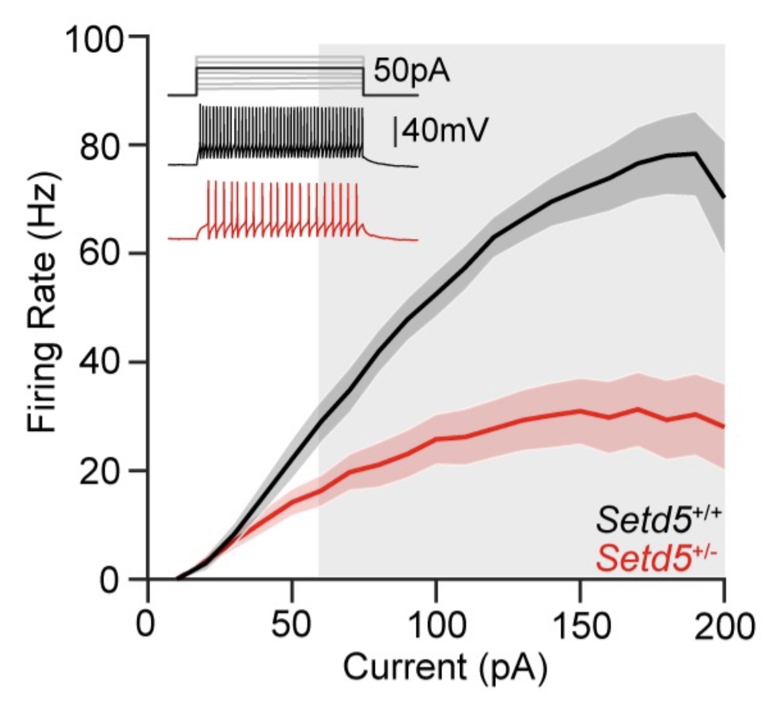
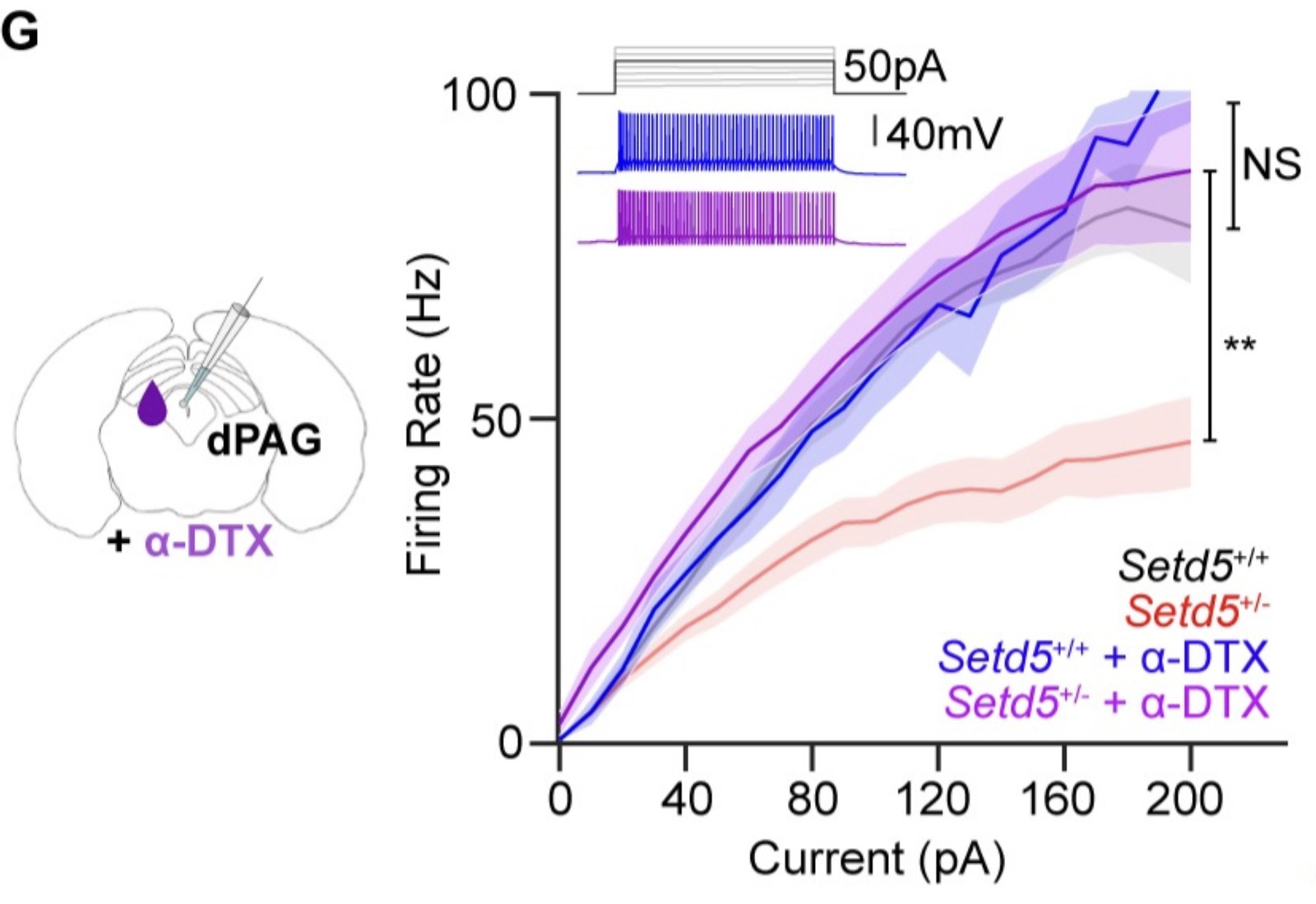
The key finding was that when we recorded from PAG cells in Setd5+/- mice, these cells were found to be hypoexcitable compared to Setd5+/+ cells. When higher and higher levels of current were injected into these cells, they were unable to generate corresponding increases in action potentials, unlike the Setd5+/+ cells. By using the drug (α-dendrotoxin) that selectively blocks Kv1 potassium channels we were able to rescue this reduced excitability phenotype which led us to conclude that overactive Kv1 channels in PAG cells were responsible for the delayed escape responses we observed in Setd5+/- mice.
As a final experiment, we implanted a cannula above the PAG of these animals and administered (α-dendrotoxin) while the mice were exposed to looming stimuli. The drug application restored normal escape timing and normal threat avoidance, showing the effect was reversible in adulthood rather than fixed early in development.
I also tested two other genetically distinct ASD mouse models (Cul3 and Ptchd1) that showed similar arrested behavioural responses to the looming stimulus, suggesting a shared “downstream” behavioural outcome despite different molecular causes.
Outcome
We found that mice with a mutation in Setd5 (a gene strongly linked to human ASD/intellectual disability) took longer to initiate escape from a looming threat and showed weaker learned avoidance of the danger zone, even though their basic vision and motor ability were normal.
In Setd5 mice specifically, we traced the problem to reduced excitability of neurons in the dorsal periaqueductal grey (dPAG), a brainstem region that triggers escape behaviour — caused by overactive Kv1 potassium channels rather than a change in how many channels were present.
Colloquial context
In truth, this project came about through the fusion of my interest and prior knowledge in the field of neurodevelopment, my burgeoning curiosity and appreciation for the tools used in systems neuroscience and the elegence and experimental utility of the neural circuit known to drive the innate, visually-driven, looming-evoked escape response in mice (Evans et al. 2018).
I had been reading some literature that described the differences in attention and visual processing often observed in autistic individuals, as well as the seminal paper referenced above that described the experimentally tractable and behaviourally robust visual escape paradigm. I had recently completed a lab rotation in the Novarino group at ISTA that had a number of genetic mouse models of autism and intellectual disability. I was intrigued as to whether we would be able to see any behavioural differences in the responses of these mice compared to their “wild type” siblings in response to this assay.
Thankfully, both my supervisor, Prof. Maximilian Joesch, and Prof. Gaia Novarino allowed me to test my hypothesis. I was given one litter of Setd5 mice, and one by one, recorded their behavioural responses to this visual loom stimulus. I tested the animals blind, meaning that I was unaware during the experiments of which animals carried the mutation in Setd5 and which didn’t. However, already having tested just 6 animals, I felt confident that there was a real difference in how some of the animals responded. While some rapidly rushed back to their shelter immediately at the onset of the loom, others displayed a short, but noticeable pause before initiating an escape of their own. When I received the genotypes of the mice, I could verify that this arresting behaviour was observed in the mice carrying the mutation. I rushed into my supervisor’s office, plots in hand, and these results then had a cascading effect on my future experiments that culminated in my PhD thesis.
Links
References
- Evans DA, Stempel AV, Vale R, Ruehle S, Lefler Y, Branco T. A synaptic threshold mechanism for computing escape decisions. Nature 558, 590–594 (2018) — the drift-diffusion model in Figure 4.
Acknowledgments
This project was carried out while I was a graduate student in Prof Maximilian Joesch’s group at the Institute of Science and Technology Austria (ISTA). I learnt a lot about coding and data science by working with Dr Olga Symonova, Dr Felipe Fredes taught me how to do the stereotaxic surgeries, Dr Tomás Vega-Zuñiga taught me how to do the silicon probe recordings. I mentored a number of rotating graduate and undergraduate summer students over the course of this project: Heloisa Chiossi, Chiara Roth, Daria Bocharova, Julie Murmann, Matteo Barel & Dafna Ljubotina - I am very grateful for all of their help. Prof Gaia Novarino provided the Setd5, Cul3 and Ptchd1 mice and support throughout. My PhD committee provided regular support: my internal committee member was Prof Ryuichi Shigemoto and my external committee member was Prof Mark Huebener.